3D Dental Scanning in Spring, TX
Clear diagnosis depends on precise imaging. When evaluating tooth position, restoration margins, or bite alignment, the dentist captures a digital intraoral scan to examine structural details in depth. For patients considering a 3D dental scan in Spring, TX, this method supports careful assessment without the need for traditional impression materials.

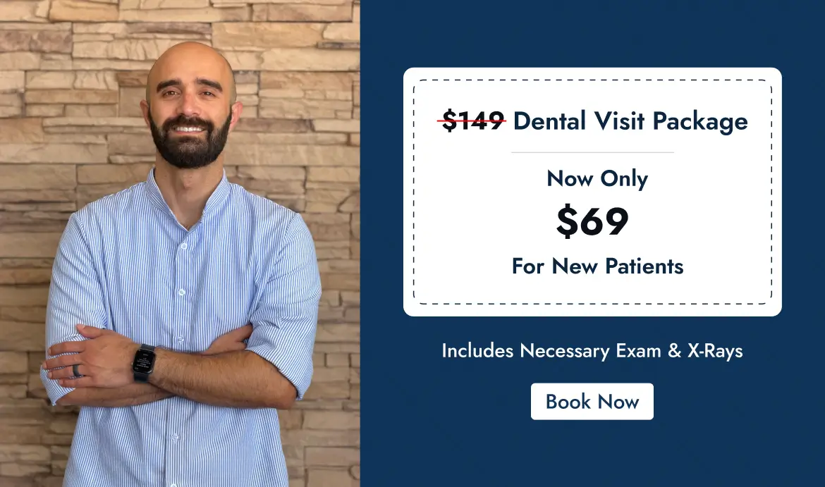
$149 Dental Visit Package
Now Only $69 for New Patients
Includes Necessary Exam & X-Rays
Book an Appointment
Fix Your Smile Today
Digital capture supports diagnosis, restorative planning, and long-term monitoring while improving comfort during the recording process.
How Digital Scanning Assists Clinical Evaluation
When a patient presents with worn enamel, a fractured restoration, spacing concerns, or bite irregularities, the dentist must first examine structural relationships. A handheld intraoral scanner records surface data and converts it into a high-resolution model.
Our dentist evaluates:
- Occlusal contact patterns
- Marginal integrity of existing crowns or fillings
- Gingival contours
- Interproximal areas are not easily visible during routine examination.
- Alignment changes over time.
This information helps plan restorations that fit precisely and function predictably.
Patients comparing options with a dentist in Spring often ask whether digital scans improve accuracy. Detailed visualization supports more controlled restorative preparation and laboratory communication.
Reducing Distortion and Retakes
Traditional impressions rely on physical material that may distort or require repetition. Digital models eliminate tray-based impressions and allow immediate verification of accuracy before laboratory submission.
For individuals evaluating dental care in North Houston, reduced remake rates and consistent marginal fit are practical considerations when selecting a provider.
Application in Restorative and Orthodontic Care
Digital scans are used when the dentist plans:
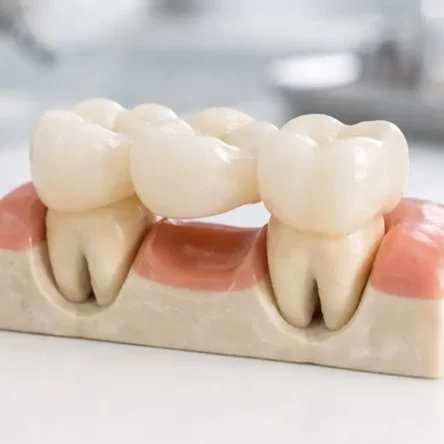
- Crown or bridge placement
- Clear aligner therapy
- Occlusal guards
- Implant-supported restorations
In each situation, Dr. Atheer evaluates the digital model to determine fit, spacing, and functional relationships before proceeding. The scan does not replace clinical examination; it supports decision-making.
Patients who search for a dentist near me frequently prioritize efficiency, but diagnostic clarity remains the primary objective.
Monitoring Structural Changes
Digital files can be stored and compared during follow-up visits. The dentist monitors:
- Progressive wear
- Minor positional movement
- Gingival recession
- Changes in occlusion
Patients living near Kuykendahl Road, Pinelake Blvd, or Eagle Bend Drive often value proximity when follow-up assessment is required.
Imaging Considerations
Intraoral scanning uses optical technology and does not involve ionizing radiation. When bone assessment or internal pathology evaluation is necessary, radiographs may still be indicated. The dentist determines which imaging method is appropriate based on clinical findings.
For those researching North Houston digital dental scanning, it is important to understand that scanning supports evaluation of surface anatomy and restorative precision rather than replacing radiographic diagnosis.
Houston Cosmetic & Family Dental integrates digital scanning into the routine diagnostic workflow to support structured planning and predictable restorative outcomes.
Frequently Asked Questions
The scanner rests lightly against the tooth surface while capturing images. Most patients tolerate the process without difficulty.
No. Radiographs evaluate bone and internal structures. Scanning documents for external tooth and soft-tissue surfaces.
Most full-arch recordings are completed within several minutes, depending on case complexity.
Precise surface capture enables the dentist to carefully assess margins and accurately communicate measurements to the laboratory.
Digital models are stored and may be compared during future examinations to monitor changes.
Problems We Treat
-
 General Dental Concerns & Preventive Needs
-
Stained or Discolored Teeth
-
Multiple Missing or Damaged Teeth
-
Cavities & Minor Tooth Damage
-
Dental Injuries & Sudden Tooth Pain
-
Crooked Teeth & Bite Misalignment
-
Gum Infections & Advanced Periodontitis
-
Early Signs of Oral Cancer
-
Loose or Damaged Dentures
-
Minor Chips & Cracks in Teeth
-
Missing Teeth & Jawbone Loss
-
Gaps from Missing Teeth
-
Severe Dental Fear or Anxiety

Other Services